Neuroimaging Research Program
The Neuroimaging Research Program within the Department of Radiology is a source of comprehensive neuroimaging research and advanced neuroimaging clinical services such as fMRI, DTI, and more, for the entire neuroimaging community at the University of Wisconsin–Madison. This program provides access to a wide variety of current modalities available within the Department of Radiology and at WIMR (MRI, PET, CT, ultrasound, fluoro) as well as in-development modalities and resources (high-density EEG, transcranial magnetic stimulation/transcranial direct current stimulation, portable neural stimulation, brain-computer interface-EEG, and mock scanners). These resources, along with simultaneous multimodal imaging (simultaneous PET-MR, PET-CT, fMRI-TMS-EEG), are available for neuroimaging use by both novice and experienced neuroimaging researchers. We augment this with full bioinstrumentation and stimulus delivery capabilities (visual/audio fMRI delivery systems, response box, physiological recording instrumentation) and provide full support in terms of protocol development/implementation, data acquisition, analysis, and storage. The Neuroimaging Research Program is distinguished from similar programs by:
- Providing the multiple imaging modalities outlined above for human and animal neuroimaging research.
- Utilizing Department of Radiology and WIMR imaging resources and having access to the hospital and clinical inpatient and outpatient populations for neuroimaging research and clinical use.
- Involving core personnel in the Departments of Radiology, Neuroradiology, and Medical Physics with expertise in various aspects of neuroimaging and collaborating with various researchers and clinicians in other departments.
The Neuroimaging Research Program is comprised of various core investigators and personnel working in the three areas outlined below. We will continue to foster additional projects in the Neuroimaging Research Program and work with external and new investigators in implementing their neuroimaging projects.
Research Areas
Learn more about the Neuroimaging Research Group at UW
Personnel
Faculty
Scientific Staff
Research Associates
Research Assistants
Staff
Students
Intradepartmental Collaborators
Interdepartmental Collaborators
Andrew Alexander, PhD Professor (Tenure) Department of Medical Physics T135 Waisman Center (608) 265-8233 alalexander2@wisc.edu
Barbara Bendlin, PhD Associate Professor, Department of Medicine bbb@medicine.wisc.edu
Rasmus Birn, PhD Associate Professor (Tenure) Departments of Psychiatry, Medical Physics 1129 WIMR I (608) 265-5609 rbirn@wisc.edu
Alison Brooks, MD, MPH Associate Professor, Department of Orthopedics and Rehabilitation (608) 263-6477 brooks@ortho.wisc.edu
Kristin Caldera
Guang-Hong Chen, PhD Professor Director of UW CT Lab Department of Medical Physics 1167 WIMR I (608) 263-0089 GChen7@wisc.edu
Dorothy Edwards, PhD
Bruce Hermann, PhD Professor Department of Neurology (608) 263-5430 hermann@neurology.wisc.edu
Hrissanthi Ikonomidou, MD Professor Department of Neurology (608) 263-5421 ikonomidou@neurology.wisc.edu
Sterling Johnson, PhD Professor, Department of Medicine (608) 256-1901 ext.11946 scj@medicine.wisc.edu
Elizabeth Meyerand, PhD Professor (Tenure) Departments of Biomedical Engineering, Medical Physics 1129 WIMR I (608) 263-1685 memeyerand@wisc.edu
Emma L. Mohr, MD, PhD Assistant Professor UWSMPH Department of Pediatrics, H6/538 Clinical Science Center(608) 265-5107 emohr@pediatrics.wisc.edu
David Noyce
David O’Connor, PhD UW Medical Foundation Professor of Pathology & Laboratory Medicine, 585 Science Drive, Madison, WI 53711(608) 890-0845 emohr@pediatrics.wisc.edu
Joanne Robbins, PhD Professor Department of Medicine (608) 280-7000 jrobbin2@wisc.edu
Robert Sanders, MD, PhD Department of Anesthesiology (608) 265-0588 rsanders4@wisc.edu
Justin Sattin, MD
Vikas Singh, PhD Professor Departments of Biostatistics and Computer Science, 5975 MSC (608) 262-8875 vsingh@biostat.wisc.edu
Justin Williams Peter Tong Department Chair and Vilas Distinguished Achievement Professor, Department of Biomedical Engineering jwilliams2@wisc.edu
Offsite Collaborators
Jeffrey Binder, MD Professor Medical College of Wisconsin Department of Neurology jbinder@mcw.edu
Bharat Biswal, PhD Professor and BME Chair New Jersey Institute of Technology Department of Biomedical Engineering bharat.biswal@njit.edu
Edgar DeYoe, PhD Professor Medical College of Wisconsin Department of Radiology deyoe@mcw.edu
John Gabrieli, PhD Professor Massachusetts Institute of Technology Department of Brain and Cognitive Sciences gabrieli@mit.edu
Argye Hillis, MD Professor Johns Hopkins University Department of Physical Medicine argye@jhmi.edu
Michael McCrea, PhD Professor Medical College of Wisconsin Department of Neurology
Allyson Rosen, PhD Clinical Associate Professor Stanford University Department of Psychiatry and Behavioral Sciences
Bart Rypma, PhD Professor University of Texas at Dallas Department of Psychiatry bart.rypma@utdallas.edu
Noam Sobel, PhD Professor Weizmann University Department of Neurobiology noam.sobel@weizmann.ac.il
Wei Zhou, MD Associate Professor Stanford University Department of Surgery weizhou@stanford.edu
Alumni
Allison Abellaneda
Ahmad Amer Abdl-Haleem
Matt Andreoli
Benjamin Austin
Dovile Baniulis
Erik Bjorklund
Kaitlin Brendel
Kirk Brown
Daniel Chu
Alexander Conway
Cole Cook
Joe Decker
Alok Desphande
Kayla Diffee
Robert Friedel
Wolfgang Gaggl, PhD
Thomas Gallagher, MD
Jeffery Garcia
Camille Garcia-Ramos
Ryan Gentile
Shawna Gloe
Max Greenstein
Scott Grogan
Jaya Gupta
Emily Hatfield
Cameron Hays
Ryan Holdsworth, MD
Natalie Htet
Gyujoon Hwang
Jessica Jiminez
Eliza K Kapinski
Rico Masodkar
Mohsen Mazrooyi Sebdani
Kanak Ryan Kimmel
Gregory Kirk
Christopher Kleefish
Nicole Komer
Arman Kulkarni
Bornali Kundu
Christian La Victoria
Aejin Kwak
Liao Sofia Linsenmeyer
Nicole Martens
Mattheiu Mayer
Ifeanyi Mbah
Matthew McMillan
Timothy Meier
Erin Meyer
Adam Milch
Rosaleena Mohanty
Anna Moldysz
Raghuvardhan Moola
Chad Moritz
Pouria Mossahebi
Krishna Mylavarapu
Tanvi Nadkami
Lin Naing
Zack Nigosyan
Oreoluwa Omoba
Joshua Pankratz
Shalini Patro
Amy Penwarden
Molly Peterson
Shruti Rajan
Ryan Raut
Rebecca Ray, PhD
Michael Regner
Peter Reiter
Sean Riley
Cameron A Rivera
Hector Salazar
Sirisha Sanamandra, MD
Naga Saranya
Addepally Paige Schultz
Lauren Seidl
Liang Shang
Anita Sinha
Hillary Smith
Julie Stamm, PhD
Joshua Suhonen
Yohan Sumathipala
Yiyou Sun
Maggie Sundstrom
Benjamin Swan, MD
Andrew Tan
Michael Turek
Amy Utter
Jed Voss
Siarhei Vysotski
Stephen Welch
Leroy Williams
Joel Wood
Hanefi Yildrim
Peng Yin
Brittany Young, PhD
Hongwu Zeng, MD
Neuroimaging Research Areas: Clinical Neuroimaging
Projects in this area are concerned with the application of advanced imaging and analysis tools for the following clinical work, which are clinically reimbursable.
- Presurgical mapping of brain tumor, epilepsy, and vascular lesions patient populations (Vivek Prabhakaran, Aaron Field, Veena Nair, and other Neuroradiology Section faculty)
- Seizure localization in Epilepsy patients utilizing simultaneous fMRI-EEG (Vivek Prabhakaran, Howard Rowley, Rama Maganti (Epilepsy Chief), Aaron Struck, and other epileptologists)
- Morphometrics (Cortical thickness, Lesion segmentation) using Neuroquant for Alzheimer’s, Epilepsy, Multiple Sclerosis, ‘chemo’ brain patients (Vivek Prabhakaran, Aaron Field, Veena Nair, and other Neuroradiology Faculty)
Neuroimaging Research Areas: Translational & Basic Science Neuroimaging
Projects in this area are concerned with the application of advanced imaging and analysis tools to investigate the neural bases of brain disorders. They focus on various diseases modeled in animals and human patient populations, along with normal animal and human investigation of sensory, affective, and cognitive processes.
- Animal to Human translational work
- Schizophrenia Model in Rodents (JP Yu)
- Stroke Work in Animal Model (Charles Strothers)
- Nonhuman Primates Model of Working Memory/Attention (Luis Populin*)
- Nonhuman Primates Model of Effect of Anesthesia on the Brain (Chris Ikonomidou)
- Disease connectome biomarkers for brain-behavioral correlations, prognostics, and tracking changes in various diseases:
- Stroke (Vivek Prabhakaran)
- Pediatric Epilepsy (Bruce Hermann*)
- Adult Epilepsy Connectome (Beth Meyerand , Bruce Hermann, Vivek Prabhakaran)
- Traumatic Brain Injury (Alison Brooks*, Mitch Tyler*)
- Alzheimer’s Disease (Sterling Johnson, Barbara Bendlin, Vivek Prabhakaran)
- Multiple Sclerosis (Aaron Field)
- Delirium (Robert Sanders*)
- Spine Imaging (Victor Haughton, Alison Grayev)
- Swallowing studies in various patient populations (JoAnne Robbins*)
- Inflammatory Bowel Disease & Cognition (Sumona Saha*)
- Carotid studies using Ultrasound & Cognition (Dempsey*, Tomy Varghese)
- Brain Tumor Diagnosis/Rx using Tumor Targeting Agent (Lance Hall, Weichert, Kuo)
*external investigators
Neuroimaging Research Areas: Neuroimaging Methods and Analysis
Projects in this area focus on increasing the speed of imaging acquisition, improving the spatial coverage of these imaging sequences, and developing innovative acquisition methods to enhance the spatial and temporal characteristics of anatomical, functional, vascular and metabolic brain signals. They are also concerned with improving the quantification of structural and functional brain images. Innovative software and algorithms are continuously being developed for use in brain segmentation, volume and cortical thickness determination, functional signal localization, as well as automated image processing and analysis pipelines.
- MRI Pulse Sequence Development (Frank Korosec, Karl Vigen)
- Multimodal Imaging (fMRI, TMS, EEG) (Vivek Prabhakaran, Veena Nair)
- Quantitative Neuroimaging Approaches (Vivek Prabhakaran, Aaron Field, Alexei Samsonov)
- Image-guided Delivery Systems (Wally Block)
- Vascular Imaging (Patrick Turski, Charles Mistretta, Kevin Johnson)
- Perfusion Imaging (CT – Guang-Hong Chen , MRI & CT – Howard Rowley)
Core Lab: Human Brain Mapping in Normal and Patient Populations
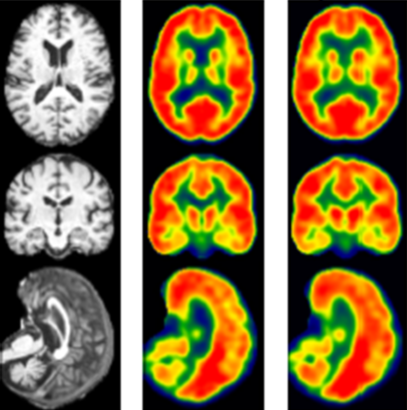
The Neuroimaging Research Core Lab is interested in utilizing multimodal imaging tools incorporating fMRI, DTI, perfusion imaging, cortical thinning, and other advanced MR imaging tools to explore various questions at the basic science, translational, and clinical levels.
The lab specializes in research on behavior, cognition and emotion; neurobiology of disease; development, plasticity and repair; and neuronal circuits.
Basic Science Level
We are currently developing cognitive models of memory and reasoning, mapping neural substrates involved in cognitive processes such as working memory and reasoning, and validating these brain maps using patient populations such as stroke patients.
Translational
The laboratory is involved in characterizing brain plasticity changes in aging as well as in various patient populations such as Stroke/Vascular lesions, Brain tumor, Epilepsy, Alzheimer’s disease, Traumatic brain injury, as well as developing novel interventions toward recovery.
Specifically, the lab combines neuroimaging measures such as functional magnetic resonance imaging (fMRI), diffusion tensor imaging (DTI), and other advanced neuroimaging as well as behavioral measures to identify prognostic factors that predict functional recovery, identify adaptive and maladaptive networks that contribute to functional recovery, and identify a critical time window for intervention in these patients. Our lab, in collaborations with Justin Williams’ Lab and TCNL, is developing Brain-Computer Interface (BCI) as well as cranial nerve noninvasive neuromodulation technologies as rehabilitation treatments for patients, which will lead to faster and more optimal levels of recovery.
Clinical
fMRI/DTI is an exciting tool that is being developed for clinical use. fMRI and DTI are used for pre-surgical brain mapping prior to the resection of brain tumors, vascular lesions, and epilepsy surgery. Brain mapping identifies functional structures involved in language, memory, vision, and sensorimotor processes to avoid during treatment. The Neuroimaging Research Core Lab is interested in further developing these tools for clinical use along with FMRI/EEG for seizure localization and FMRI/TMS for validation of brain mapping.