Sandip Biswal investigates new method to pinpoint pain

Picture people in small towns and big cities across the United States holding up flashlights. A satellite image taken at night would show clusters of light brightly illuminating densely populated areas and flickering faintly in less crowded zones.
A viewer could quickly identify where major metropolitan areas like Chicago, New York City, and Los Angeles are located. This is analogous to Sandip Biswal, MD’s work identifying clusters of pain receptors in the body using alternative imaging techniques.
“In a sense, that’s what I’m trying to do by combining positron emission tomography (PET) and magnetic resonance imaging (MRI),” Dr. Biswal said. “I want to pinpoint exactly where there is a high density of pain receptors or painful inflammatory activity. Doing so can give us insights to the precise location where your pain arises and thereby provide more accurate information to help manage your pain. ”
Dr. Biswal has been studying musculoskeletal pain since the late 1990s. Pain is one of the most common reasons to seek medical care and can be physically and mentally debilitating. Despite its prevalence, pain is often misunderstood and poorly treated.
During medical school, Dr. Biswal noticed how pain patients were mismanaged.
“It kind of broke my heart because I’ve had friends, including relatives and my mom, who have had bouts with chronic pain or pain for reasons that were not entirely clear,” he said. “I saw them go through a battery of clinic visits and tests and not get really any successful help or therapy.”
Figuring out how to be better diagnosticians for people in pain motivated Dr. Biswal’s decision to pursue radiology. But he realized that the methods available did not give him enough information.
‘Little light beacons’
MRI is the workhorse for showing the body’s anatomy in exquisite detail, but structural abnormalities do not always show where pain is coming from. Many findings seen on MRI — such as cartilage loss, disc degeneration, or tendon changes — can be present in people with little or no pain, while some patients with severe pain may have only subtle abnormalities on conventional imaging.
In other words, MRI excels at showing what looks abnormal, but it is often less certain whether that abnormality is actually causing a patient’s pain.
“Going from individual to individual, I can’t reliably tell you if that person is in pain just based on the MRI,” Dr. Biswal said.
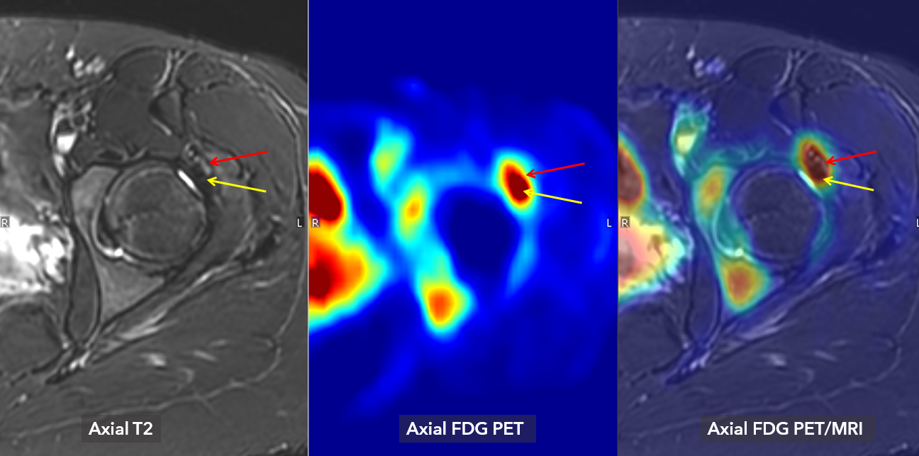
But when MR and PET imaging are used together, Dr. Biswal can create two images, fuse them, and pinpoint with “a confidence of almost two to three millimeters” where pain is being generated in a patient.
The hybrid PET/MRI approach combines the sensitivity of PET imaging and the soft tissue contrast and spatial resolution of MRI. With this technique, Dr. Biswal uses fluorodeoxyglucose (FDG) as a tracer that seeks abnormal, painful inflammatory activity. Because FDG is radioactive, it emits gamma rays — the “light beacon” — that can be detected by PET imaging.
“You inject this drug that now has this radiolabeled positron emitter on it, and it swims around your whole body looking for that receptor or that painful inflammatory activity,” Dr. Biswal explained. “If you have a lot of receptors or painful inflammatory activity at one site, these drugs with the little light beacons will cluster at that site.”
Then, an MRI is used to identify where the clusters of light are located, for example, in a specific muscle, nerve, joint, or ligament. After analyzing them together, Dr. Biswal can recommend an image-guided injection to confirm the area is painful or an injection of lidocaine and a steroid for symptomatic relief. If the injection proves to relieve their pain, that helps confirm the location of the pain generator and inform the next step, which could be an additional injection or surgery.
Promising results
This strategy is promising. In the 2025 publication “Impact of FDG PET/MRI on Management of Patients with Chronic Spinal Pain,” Dr. Biswal and the co-authors found that this method “demonstrated an overall positive impact on management of patients with chronic spinal pain, mainly achieved by identifying previously unknown foci of pain generation and enabling targeted interventions.”
A 2024 study of 12 people with clinically challenging spinal or lower extremity pain found that using hybrid PET/MRI resulted in a new procedure or increased confidence of subsequent management decisions. This study found that the hybrid imaging method “can be helpful in identifying potential pain generators that were otherwise challenging to reveal and is preliminarily altering management or improving diagnostic confidence in providing favorable outcomes to individuals suffering from chronic pain.”
In 2026, Dr. Biswal and others, including Ali Pirasteh, MD and Kenneth Lee, MD, MBA, co-authored “18F-FDG PET/MRI Identifies Previously Unrecognized Pain Generators in Patients with Persistent Pain After Hip Surgery.” They concluded that this method is a “clinically useful adjunct for evaluating persistent post-arthroscopic hip pain.”
When a patient suffers from chronic pain, a diagnosis can be affirming even if the pain isn’t cured.
“We definitely get the patient who’s been frustrated or the referring physician who’s been frustrated for not being able to achieve a level of comfort for that patient,” Dr. Biswal said. “You can see how devastating this whole thing has been for them and to offer them some sort of sanity and relief, even just with the diagnosis, has been really rewarding.”
Future focus
Looking to the future, Dr. Biswal wants to develop a tracer that is more specific than the one currently used and could also be used as a medication for pain, which would open diagnostic and therapeutic opportunities. He is working closely with Aaron Lebeau, PhD; Kendall Barrett, PhD; Jonathan Engle, PhD; and Jason Mixdorf, PhD to develop the next generation of pain imaging tracers.
He is also researching how artificial intelligence tools could decrease the amount of time it takes to read these complex scans.
To make a “sizeable contribution” to pain research, Dr. Biswal said he hopes to convince others to work with him. He began working closely with Rianne van der Heijden, MD, PhD, who is based out of Erasmus University Medical Center in the Netherlands, when she came to UW–Madison as a Bracco Fellow in 2022. Now, Dr. van der Heijden has a team of people reading these types of scans. Dr. van der Heijden received research and development funding from the department to continue studying how innovative functional imaging can increase understanding of chronic pain.
A challenge in recruiting other researchers is that his work requires proficiency in musculoskeletal radiology or neuroradiology in addition to reading PET scans, which is traditionally taught through a nuclear medicine curriculum. This was the case for Dr. Biswal who made it his mission to become fluent in this alternate imaging technique.
“I want to leave this world thinking we helped people suffering from pain,” Dr. Biswal said.
This work also requires a fair amount of sophisticated equipment, multidisciplinary expertise, and infrastructure that includes cyclotrons, radiochemists, PET/MRI scanner, nuclear medicine physicians, synthetic biologists, pain practitioners, and surgeons.
“I am very fortunate to be here at UW–Madison since it is one of the few places in the country that can provide the support needed and the right environment for this work,” Dr. Biswal said.