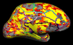
Energy Estimates rendered on Cortical Surface
The Neuroimaging Research Core Lab is interested in utilizing multimodal imaging tools incorporating fMRI, DTI, perfusion imaging, cortical thinning, and other advanced MR imaging tools to explore various questions at the basic science, translational, and clinical levels.
The lab specializes in research on behavior, cognition and emotion; neurobiology of disease; development, plasticity and repair; and neuronal circuits.
Basic Science Level
We are currently developing cognitive models of memory and reasoning, mapping neural substrates involved in cognitive processes such as working memory and reasoning, and validating these brain maps using patient populations such as stroke patients.
Translational
The laboratory is involved in characterizing brain plasticity changes in aging as well as in various patient populations such as Stroke/Vascular lesions, Brain tumor, Epilepsy, Alzheimer’s disease, Traumatic brain injury, as well as developing novel interventions toward recovery.
Specifically, the lab combines neuroimaging measures such as functional magnetic resonance imaging (fMRI), diffusion tensor imaging (DTI), and other advanced neuroimaging as well as behavioral measures to identify prognostic factors that predict functional recovery, identify adaptive and maladaptive networks that contribute to functional recovery, and identify a critical time window for intervention in these patients. Our lab, in collaborations with Justin Williams’ Lab and TCNL, is developing Brain-Computer Interface (BCI) as well as cranial nerve noninvasive neuromodulation technologies as rehabilitation treatments for patients, which will lead to faster and more optimal levels of recovery.
Clinical
fMRI/DTI is an exciting tool that is being developed for clinical use. fMRI and DTI are used for pre-surgical brain mapping prior to the resection of brain tumors, vascular lesions, and epilepsy surgery. Brain mapping identifies functional structures involved in language, memory, vision, and sensorimotor processes to avoid during treatment. The Neuroimaging Research Core Lab is interested in further developing these tools for clinical use along with FMRI/EEG for seizure localization and FMRI/TMS for validation of brain mapping.